Back in April, when I first learned of my mutation, my gynecologist immediately scheduled me for my first ever breast MRI. However, I had to delay it because I had just received my second COVID vaccine. The vaccine sometimes results in swollen lymph nodes which can be mistaken for a mass on a breast MRI, so I was advised to wait 4-6 weeks.
I went for the MRI on May 17. I was notified the next day that they “saw something,” and I was scheduled for an ultrasound the following week. My doctor and the radiologist assured me that it is common to find these small masses on an MRI, especially the first time; this test is much more sensitive than a mammogram.
I had the breast ultrasound on May 25. The technician could not find the mass seen on the MRI. Although the radiologist and my doctor continued with their assurances, they scheduled me for a biopsy for later that week.
The word “biopsy” is scary. I had never had one before. In addition, I was able to get the first 2 procedures done at an outpatient imaging clinic that had just opened in town, affiliated with the hospital where I had been having mammograms for years. It was bright, clean, and intimate, since not a lot of people know about it. I could be in and out in an hour. By contrast, the MRI-guided biopsy had to be done at the hospital. I blocked off 2½ hours for the procedure.
MRIs in general have never bothered me. I haven’t had many, but I have always been able to tune out the noise and banging, close my eyes, and relax. I expected similar this time.
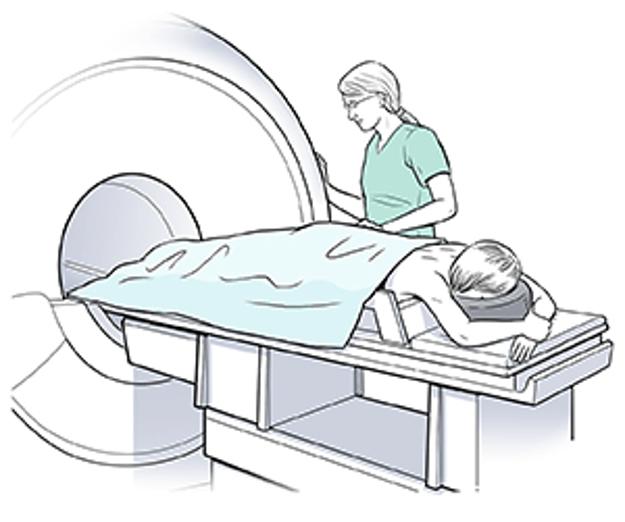
In a core needle MRI guided biopsy, you first lie in an MRI machine on your stomach with your breasts hanging down and your arms outstretched. Doesn’t sound very comfortable, does it? It’s not nearly as easy to close your eyes and relax when you’re lying face down with your arms outstretched.
In an MRI-guided biopsy, contrast material is administered via IV, to better visualize the mass. The radiologist uses the MRI to map out a grid of the breast, and then plots the mass on the grid. Once the mass is plotted, the radiologist calculates the position and depth the needle must be inserted to access the mass.
Under a local anesthesia, the needle is inserted to the location of the mass, using the MRI to verify its position. Then a cluster of cells is removed through the needle. After the needle is withdrawn, a guide or clip is placed at the site of the biopsy for future reference. At that point the MRI is over, but then a mammogram is performed to confirm the correct location of the clip.
Of course, you have to stay very still through all of this. The worst part is the feeling of numbness/stiffness that inevitably occurs after lying in this uncomfortable position for an extended period of time (the nurse and technician massaged my arm, shoulder, and neck to try to keep the circulation going).
The medical personnel did a great job of explaining to me what would happen. The biggest surprise was how tired I was afterwards. My procedure was scheduled for 9AM; I had expected to go back to work in the afternoon. However, I was so exhausted afterwards that I texted my boss and told her I wouldn’t be working that afternoon. I went home and took a long nap.
A few days later the results came in—benign. No cancer cells. I was relieved, but also not surprised. I guess I still didn’t believe any of this was really happening to me, or that I could even get breast cancer. That happens to someone else, right?
